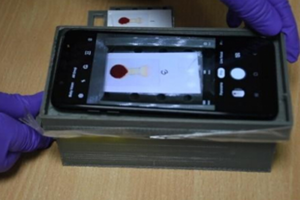
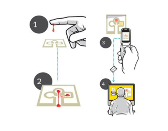
Portable and thus spot diagnosis is possible
Functional with limited resources
Easy handling
Mass fabrication is possible
Inexpensive
Involvement of minimal infrastructures
Rapid and easy monitoring
Automated
Printed Paper Channels for Blood Pathology
Lifestyle factors in the underserved community
Current methods of diagnostics require sophisticated lab-based procedures
A low cost, portable, handheld imaging device to screen oral cancer and pre-cancer based on measured change in blood flow rate of the tissue fro thermal imaging and analytics.
Successful phase-I clinical trial with more than 60 patients indicated more than 96.66 % accuracy in detecting oral cancer and pre-cancer.
The early prototype developed is ready to be deployed in the field by minor sophistication and automation.
Method can be extended to other forms of cancer
“COVICUBE” –which can simultaneously measure respiration rate, Oxygen saturation, breathing rate, Temperature has been prototyped and validated in rural settings. This will provide a much-needed low-cost integrated solution for screening the rural population through the same women rural health workers.
1. Apply glycerin on the paper pad of the cartridge with paint brush. Make sure that the glycerin is completely absorbed within the paper and no surplus volume of glycerin remains on the paper surface
2. Properly align the paper cartridge and the smartphone on the designated slot of the image acquisition platform.
3. Open the camera of the smartphone and adjust the magnification to 2.5X.
4. Perform finger pricking and deposit one drop of finger pricked blood on the wet paper cartridge just by touching the blood drop on the paper surface.
5. Capture the image of the blood pattern after 3-4 secs of dispensing blood.
6. Open smartphone app ‘Hemo-app’.
7. Click on ‘Start a new test’.
8. Click on ‘Upload image from Gallery’
9. Choose the recently captured blood pattern image.
10. Wait for the result display on the smartphone app.
11. Dispense the used paper cartridge into biosafety-waste bin.
1. Start with calibrating the impedance analyzer.
2. Connect the device electrodes with the impedance analyzer.
3. Dispense a drop of blood on the strip.
4. Instantly record the impedance magnitude at desired optimal frequency.
5. Feed the obtained impedance magnitude in the smartphone app.
6. Get the Hematocrit and Hemoglobin value result in the smartphone app.
This is a deep learning-enabled android app to predict the vulnerable COVID-19 patients based on digital frontal chest X-ray images. This app is designed to individuate COVID-19 cases from no- findings and non-COVID Pneumonia. Using this application, you can get the automated classification of frontal chest X-rays revealing fine-grained variability in appearance that are not distinguishable by Pulmonologist. Here, deep convolutional neural networks (CNN) were used to accomplish highly variable tasks in many types of fine-grained objects in the images. For the development of the CNN model, the classification of chest X-rays was carried out using a single CNN with transfer learning, formed end-to-end from direct images, using only pixels and disease tags as entries. The used CNN model was trained on a dataset accessed from public repository consisting of a total of 1697 frontal X-ray images (549 samples of COVID-19, 576 samples of no findings, and 572 samples of non-COVID pneumonia) with high classification accuracy.